 the following parameters must be controlled For x-ray machines:
1.anode voltage;
2.exposure time;
3.the total thickness of the filter;
4.Kerma in the air;
5.radiation output;
6.correspondence of edges of light and radiation fields;
7.correspondence of the centers of light and radiation fields;
8.compliance of the center of the light field and the cassette;
9.x-ray beam perpendicular;
10.automatic collimation check;
11.automatic exposure control system (AUE);
12.the size of the focal spot.
For x-ray and angiographic devices, the following parameters must be controlled:
1.anode voltage;
2.power input surface dose;
3.dose rate at the input surface of the image amplifier;
4.timer;
5.the total thickness of the filter;
6.image settings (x-ray field area and x-ray image amplifier(URI), contrast and resolution of the monitor).
For fluorographic devices, the following parameters must be controlled:
1.anode voltage;
2.exposure time;
3.Kerma in air.
For mammography machines, the following parameters must be controlled:
1.anode voltage;
2.exposure time;
3.the total thickness of the filter;
4.Kerma in the air;
5.radiation output;
6.coincidence of light and radiation fields;
7.automatic exposure control system (AUE);
8.compression and compression thickness;
9.image settings (Contrast and resolution).
For CT scanners, the following parameters must be controlled:
1.image noise;
2.CT values of numbers;
3.constant CT of number;
4.computed tomographic dose index CTDI;
5.thickness of tomographic slice;
6.high contrast resolution;
7.table installation accuracy.
the following parameters must be controlled For dental devices:
1.distance to the patient's skin;
2.anode voltage;
3.exposure time;
4.Kerma in the air;
5.radiation output;
6.the thickness of the overall filtration.
Technical controls:
Over the past twenty years, numerous devices and test objects for the control of x-ray machines have been created, as well as methods for their application have been developed.
abroad controls are made by firmsPTW-FreiburgVictoreenUnfors InstrumentsRTI-Electronics and others.
To ensure efficiency and minimize labor costs for testing x-ray medical equipment, in world practice, devices with the principle of "non-invasive control" are used, i.e. do not require connection to the electrical circuit of the device.
the Principle of operation of devices for measuring parameters such as anode voltage is based on the principle of radiation measurements - determination of x-ray energy using several pairs of detectors (silicon detectors with different input filtration) and the subsequent calculation based on the programmed dependencies.
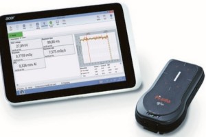
DOSIMETERUNFORS XI
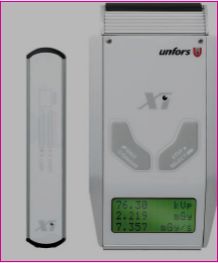 the Dosimeter can be used for measurements to a variety of medical or x-ray installations and devices. In particular:
>CT scanners,
>dental devices
> mammography devices
>various instruments for surgeries
>x-ray apparatus
Dosimeter universal for monitoring the characteristics of x-ray machines Piranha
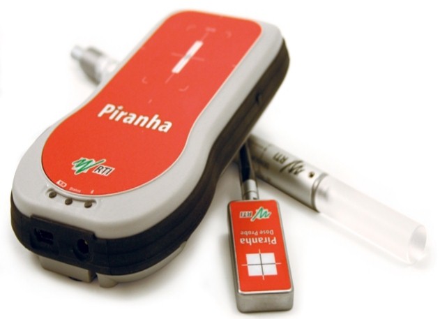
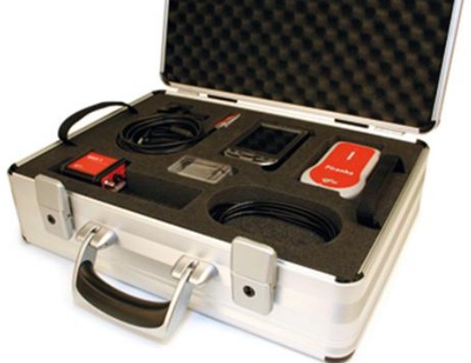
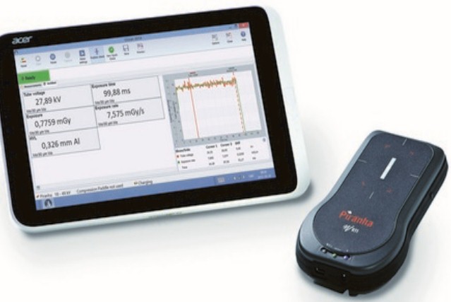
Piranha - universal x-ray dosimeter, a new generation device for monitoring the electrical and radiation parameters of medical x-ray machines.

Purpose:
-air Kerma measurement;
-Kerma power in the air;
-anode voltage on x-ray tube;
-exposure time;
-number of pulses;
-anode current;
-anode current products for exposure time;
-half attenuation layer (SPO);
-brightness;
-illumination.
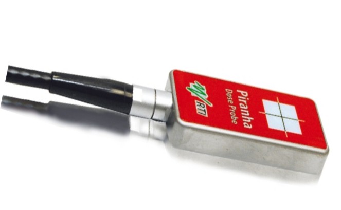
definition:
-forms of anode voltage, anode current and air Kerma power signals;
-pulse frequencies;
-pulse durations;
-air Kerma and output air Kerma per pulse;
-full filtering;
-dose profile for computed tomography (CT);
-width of the dose profile at half height (FWHM);
-CT dose indices (CTDI, CTDI100, CTDIw, CTDIvol);
-dose products per length (DLP);
-scattering index for CT.
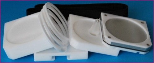
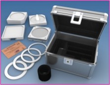
Mammographic special phantom FMCS Tissue-Equivalent Mammography Phantom 011A
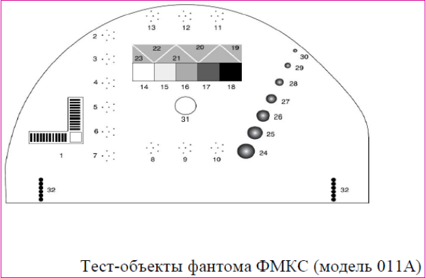 Properties:
-Phantom of tissue-equivalent material simulates a layer of compressed breast 4.5 cm thick
-phantom Test objects of various sizes, shapes, and densities simulate hardening, fibrous duct seals, and tumor-like masses
-Phantom comes with hand microscope
Features:
-option to check for any mammography system on the ability of early detection of signs of breast cancer
-phantom Test structures have a wide range of sizes, providing image control of all types of mammography devices
-Can be used for both periodic and current mammography monitoring
-Ability to simulate different thicknesses of compressed Breasts with additional tissue equivalent plates.
Properties:
-Phantom of tissue-equivalent material simulates a layer of compressed breast 4.5 cm thick
-phantom Test objects of various sizes, shapes, and densities simulate hardening, fibrous duct seals, and tumor-like masses
-Phantom comes with hand microscope
Features:
-option to check for any mammography system on the ability of early detection of signs of breast cancer
-phantom Test structures have a wide range of sizes, providing image control of all types of mammography devices
-Can be used for both periodic and current mammography monitoring
-Ability to simulate different thicknesses of compressed Breasts with additional tissue equivalent plates.compression Meter 163 The Breast Compression Test Device

Control of the correct operation of the compression device of mammographic x-ray machines.
quality Control of this parameter of mammographic x-ray systems is important for obtaining a reliable image during the diagnostic study. Mammograph breast compressor minimizes patient mobility, reduces the possibility of scattered radiation, increases image contrast and reduces radiation load on the breast by reducing the overall thickness of breast tissue. Purpose:compression Meter is used to measure the compression force (compression) in the manual and automatic modes of a specific mammographic x-ray machine.
Properties: a dynamometer is used To measure the compression force in the compression meter. The maximum compression force will be displayed until the reset button is pressed (it returns the arrow to "0").
Dental phantom kit PRO-DENTSET
Purpose: -Acceptance, periodic and ongoing tests for the constancy of the parameters of dental x-ray machines -Periodic and ongoing inspection of all types of dental x-ray machines (digital and film, intraoral, cephalometric and panoramic) to determine:
-beam alignment and collimation;
-the diameter of the beam at the outlet of the tube;
-spatial resolution;
-dose constancy;
-contrast resolution;
-constancy of the photo process quality.
Properties: universality for all types of dental x-ray machines;
modular design.
Composite CT-phantom CTDI 320/160/100
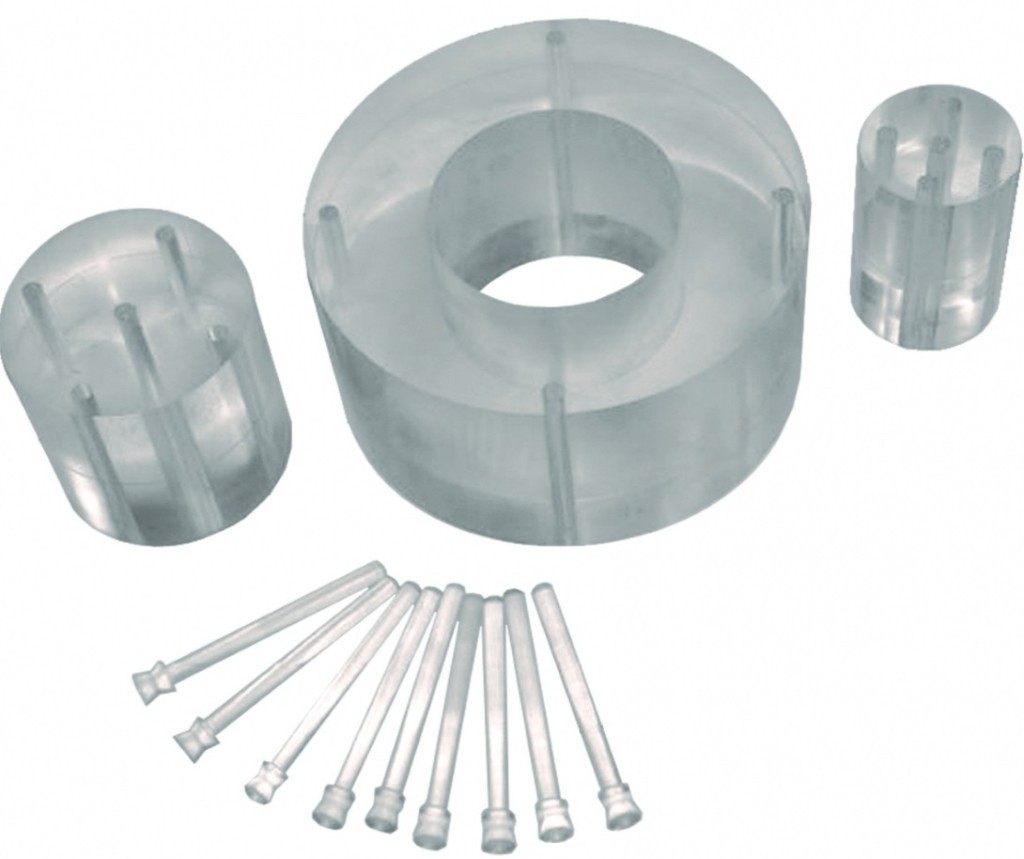
Purpose: CT phantom is designed to simulate the body (head) of the patient in determining the tomographic dose index (CTDI).
Properties
-phantom consists of three parts:
-body phantom;
-the head phantom, which is also a children's body phantom;
-children's head phantom;
-all parts of the CT phantom are made of solid acrylic (polymethylmethacrylate) with a diameter of 320, 160 and 100 m, respectively;
-each part of the CT phantom has four through holes for sensors, the holes are located through 90° in a circle and at a distance of 10 mm from the surface of the phantom. Children's head phantom has an additional hole in the center;
-the complete set of each part of the CT phantom includes rods to fill unused holes;
-a case is supplied for transportation and storage of CT phantom.
AAPM CT combined phantom for computed tomography : 610 AAPM CT Performance Phantom
Periodic and ongoing quality control of CT scanners: noise; low contrast sensitivity; accuracy of positioning; the increase of rigidity of beam; thickness of layer; spatial resolution; dependence on size; the absorbed dose (TLD); the linearity of the CT index.
Properties:
-simple control system of basic parameters of computer tomographs;
-all phantom components are mounted in a compact, transparent container;
-the possibility of modeling the increase of the stiffness of the beam in the bone;
-the container has containers of various sizes that can be filled with dextrose solution to form low-contrast test objects;
-built-in test objects of different density;
can be placed inside the phantom TLD dosimeters
ACR CT phantom for computed tomography Ref:464
Purpose:toontrol image quality of computer tomography scanners at starting, periodic and current tests.
Properties:
-phantom is made of homogeneous and stable polymer "Solid water"®;
-phantom consists of four main modules: match module; low contrast module; homogeneity module; high contrast module;
-control of all image quality indicators;
-portability and versatility;
-presence of markers "head", "legs", "top" for correct positioning.
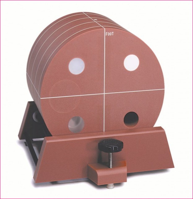
TOUR-01 universal set of geometric distortion control of General-purpose RDA images
Purpose: periodic and current control of geometric distortions of General-purpose RDA images
match fields
-x-ray beam perpendicularity;
-field size;
-image distortions;
-spatial resolution;
-contrast sensitivity.
Properties:
-simplicity and clarity of control;
-versatility and functional flexibility;
-complex control for one exposure.
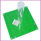
high-Precision X-Ray Pattern spatial resolution Test objects
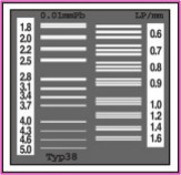
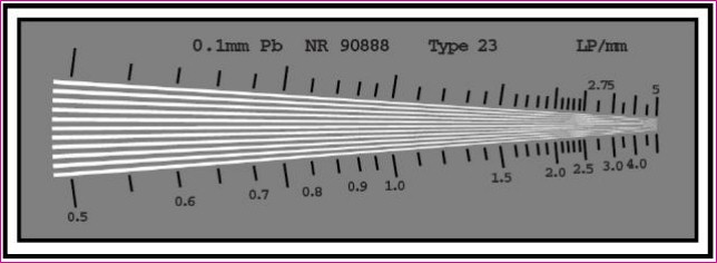
Purpose: periodic and current spatial resolution monitoring.
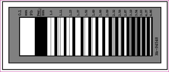
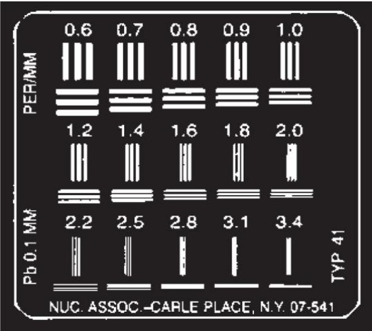
KRFA filter Set

Control of half attenuation layer (SPO) of x-ray machines of various types
Purpose:half attenuation layer control (SPO).
Properties: -ability to set any thickness required by the methods of control;
-different materials for different applications.
optical densitometer UNILIGHT D Ref:VD0204108
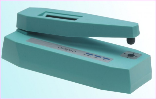
Purpose:optical density measurements of radiographs.
Properties:
-highlighted workspace;
-no time required for preheating;
-automatic health self-test;
-automatic zeroing and calibration;
-calibration radiograph is included in the basic kit.